
Topical control of wound healing by nanocerium in carcinogenesis
Авторы
-
Ekaterina V. Blinova
Sechenov University
 https://orcid.org/0000-0003-0050-0251
-
Denis N. Shimanovsky
Sechenov University
https://orcid.org/0000-0002-8211-8138
-
Ivar S. Struts
Sechenov University
https://orcid.org/0009-0001-9443-1244
-
Kirill D. Blinov
Sechenov University
https://orcid.org/0009-0002-7195-2191
-
Varvara S. Berseneva
Sechenov University
https://orcid.org/0009-0007-8972-541X
-
Evgenii V. Grebenkin
National Research Nuclear University MEPhI; Moscow Botkin Multidisciplinary Scientific-Clinical Center
https://orcid.org/0000-0002-4990-6722
-
Igor V. Lunev
National Research Nuclear University MEPhI
https://orcid.org/0009-0003-9514-0814
-
Anna V. Epishkina
National Research Nuclear University MEPhI
https://orcid.org/0000-0002-7824-7949
-
Ekaterina A. Koreeva
National Research Nuclear University MEPhI
https://orcid.org/0009-0009-4214-9974
-
Masud A. R. Farakhmand
National Research Nuclear University MEPhI
https://orcid.org/0009-0009-6828-9078
-
Sergey P. Timoshkin
National Research Nuclear University MEPhI
https://orcid.org/0000-0001-5518-6859
-
Dmirty S. Blinov
Dmitry Rogachev National Medical Research Center of Pediatric Hematology, Oncology and Immunology
https://orcid.org/0000-0002-8385-4356
-
Evgenia V. Shikh
Sechenov University
https://orcid.org/0000-0001-6589-7654
DOI:
https://doi.org/10.18413/rrpharmacology.12.1068Аннотация
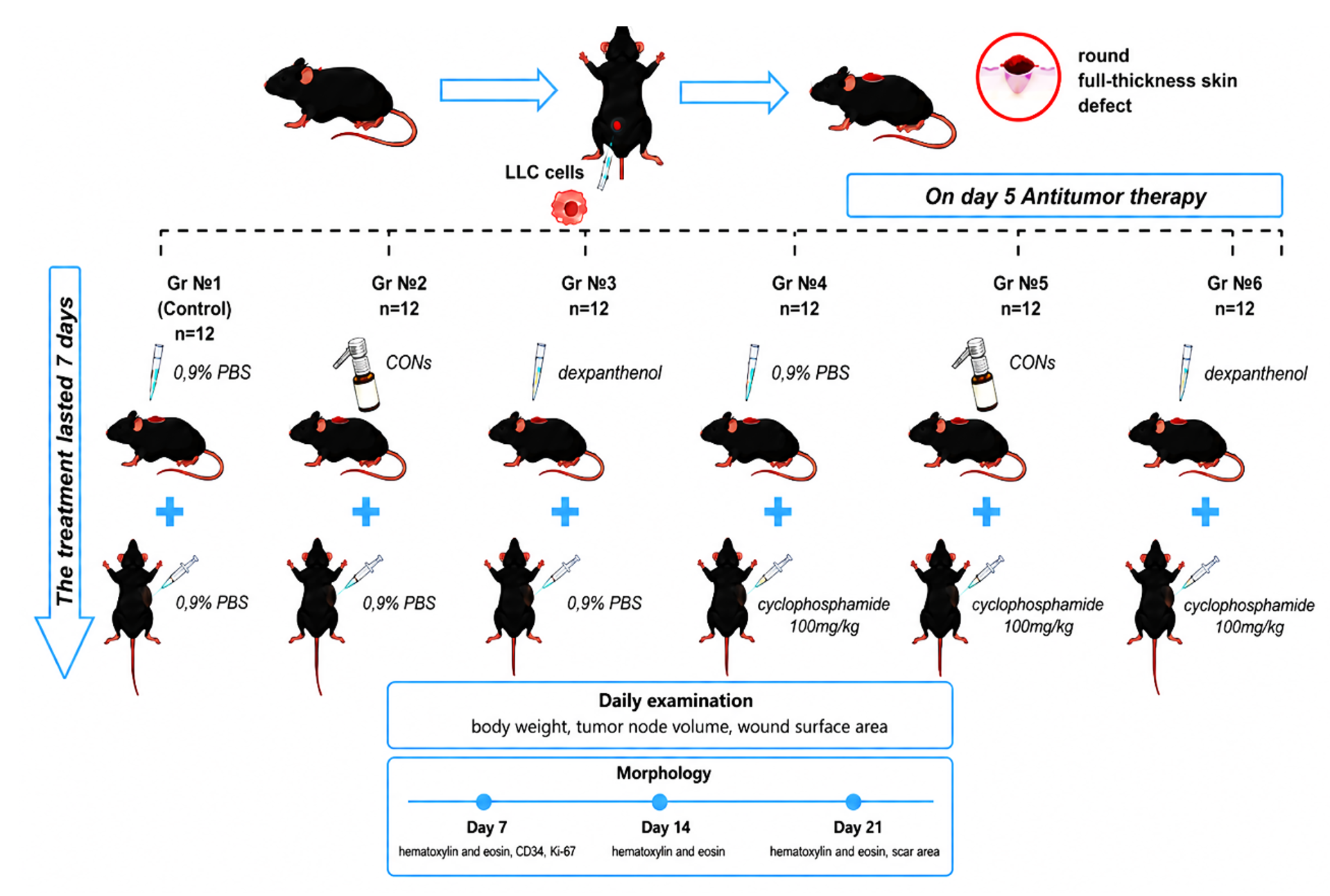
Introduction: Skin wound healing in cancer patients is a complex issue, influenced by both the tumor process itself and the effects of antitumor therapy. This study assessed the efficacy of various skin wound treatment regimens in mice with transplanted Lewis lung carcinoma (LLC).
Materials and Methods: 72 C57BL/6 mice were divided into six groups, including control, topical application of 1% cerium nanoparticles colloidal solution, 5% dexpanthenol spray, 100 mg/kg cyclophosphamide solution, and a combination of these.
Results and Discussion: It was found that topical application of cerium nanoparticles without chemotherapeutic treatment significantly shortened the time of wound regeneration to 16.5±1.0 days (p≤0.05) and reduces a postoperative scar (6±0.3 mm², p≤ 0.0001). Systemic chemotherapy treatment increased the healing time (22.5±1.0 days) and scar area (55.0±0.7 mm²), while reducing a tumor volume by 34% comparing to such in the control group. Combination therapy by dexpanthenol and cyclophosphamide promoted an antitumor effect and accelerated a tissue repair (16.3 ± 0.8 days). At the same time, local application of cerium nanoparticles did not alter regeneration (21.5 ± 1.0 days); however, the scar area was smaller (10.0 ± 0.7 mm²). Histological analysis revealed that the highest vascular density and maturity of granulation tissue in a wound were observed when treated cerium nanoparticles, while epithelial cell proliferation was reduced by cyclophosphamide.
Conclusion: The obtained data indicate the potential of topical application daily for 7 days of 1% nanocerium to accelerate wound regeneration in cancer patients without chemotherapy, and 5% dexpanthenol to accelerate skin regeneration in cancer patients receiving chemotherapy.
Графическая аннотация
Ключевые слова:
Lewis lung carcinoma, mice, nanocerium, dexpanthenol, cyclophosphamide, regeneration, wound healing gelБиблиографические ссылки
Behranvand N, Nasri F, Zolfaghari Emameh R, Khani P, Hosseini A, Garssen J, Falak R. (2022) Chemotherapy: a double-edged sword in cancer treatment. Cancer Immunology, Immunotherapy 71(3): 507–526. https://doi.org/10.1007/s00262-021-03034-y [PubMed] [PMC]
Bosari S, Lee AKC, DeLellis RA, Wiley BD, Heatley GJ, Silverman ML (1992) Microvessel quantitation and prognosis in invasive breast carcinoma. Human Pathology 23(7): 755–761. https://doi.org/10.1016/0046-8177(92)90344-3 [PubMed]
Carvalho PM, Felício MR, Santos NC, Gonçalves S, Domingues MM (2018) Application of light scattering techniques to nanoparticle characterization and development. Frontiers in Chemistry 6: 237. https://doi.org/10.3389/fchem.2018.00237 [PubMed] [PMC]
Deptuła M, Zieliński J, Wardowska A, Pikuła M (2010) Wound healing complications in oncological patients: perspectives for cellular therapy. Postȩpy Dermatologii i alergologii 36(2): 139–146. https://doi.org/10.5114/ada.2018.72585 [PubMed] [PMC]
Fadilah NIM, Phang SJ, Kamaruzaman N, Salleh A, Zawani M, Sanyal A, Maarof M, Fauzi MB (2023) Antioxidant biomaterials in cutaneous wound healing and tissue regeneration: a critical review. Antioxidants 12(4): 787. https://doi.org/10.3390/antiox12040787 [PubMed] [PMC]
Kuo PJ, Lin PC, Hsieh CH (2025) Wound healing in cancer patients under immunotherapy. International Journal of Surgery 111(10): 7087–7098. https://doi.org/10.1097/JS9.0000000000002819 [PubMed] [PMC]
Levra Levron C, Elettrico L, Duval C, Piacenti G, Proserpio V, Donati G (2025) Bridging tissue repair and epithelial carcinogenesis: epigenetic memory and field cancerization. Cell Death and Differentiation 32(1): 78–89. https://doi.org/10.1038/s41418-023-01254-6 [PubMed] [PMC]
Matsuyama K, Chiba Y, Sasaki M, Tanaka H, Muraoka R, Tanigawa N (1998) Tumor angiogenesis as a prognostic marker in operable non-small cell lung cancer. Annals of Thoracic Surgery 65(5): 1405–1409. https://doi.org/10.1016/s0003-4975(97)01416-1[PubMed]
Miller ID, Payne S, Ogston KN (2002) A new histological grading system to assess response of breast cancer to primary chemotherapy. International Journal of Oncology 20(4): 791–796.
Nelson BC, Johnson ME, Walker ML, Riley KR, Sims CM (2016) Antioxidant cerium oxide nanoparticles in biology and medicine. Antioxidants 5(2): 15. https://doi.org/10.3390/antiox5020015 [PubMed] [PMC]
Nwosu N (2024) Cancer: A disease of modern times? Cureus 16(11): e74666.https://doi.org/10.7759/cureus.74666 [PubMed] [PMC]
Payne WG, Naidu DK, Wheeler CK, Barkoe D, Mentis M, Salas RE, Smith DJ Jr, Robson MC (2008) Wound healing in patients with cancer. Eplasty 8: e9. [PubMed] [PMC]
Shekhter AB, Pekshev AV, Vagapov AB, Butenko AV, Fayzullin AL, Rudenko TG, Sharapov NA, Serejnikova NB, Vasilets VN (2020) Dose-dependent effect of plasma-chemical NO-containing gas flow on wound healing. An experimental study. Clinical Plasma Medicine 19-20: 100101. https://doi.org/10.1016/j.cpme.2020.100101
Słonimska P, Sachadyn P, Zieliński J, Skrzypski M, Pikuła M (2024) Chemotherapy-mediated complications of wound healing: An understudied side effect. Advances in Wound Care 13(4): 187–199. https://doi.org/10.1089/wound.2023.0097 [PubMed] [PMC]
Sundaram GM, Quah S, Sampath P (2018) Cancer: The dark side of wound healing. The FEBS Journal 285(24): 4516–4534. https://doi.org/10.1111/febs.14586 [PubMed]
Zawrzykraj M, Deptuła M, Kondej K, Tymińska A, Pikuła M (2023) The effect of chemotherapy and radiotherapy on stem cells and wound healing. Current perspectives and challenges for cell-based therapies. Biomédecine and Pharmacothérapie 168: 115781. https://doi.org/10.1016/j.biopha.2023.115781 [PubMed]
Загрузки
Опубликован
Как цитировать
Выпуск
Раздел
Лицензия
Copyright (c) 2026 Blinova EV, Shimanovsky DN, Struts IS, Blinov KD, Berseneva VS, Grebenkin EV, Lunyov IV, Epishkina AA, Koreeva EA, Farakhmand MAR, Timoshkin SP, Blinov DS, Shikh EV

Это произведение доступно по лицензии Creative Commons «Attribution» («Атрибуция») 4.0 Всемирная.
 Русский
 English

