
Morphometric visualization, analysis, and assessment of atherosclerotic lesions in an apolipoprotein E–deficient mouse model (ApoE−/−)
Авторы
-
Петр Р. Лебедев
Белгородский государственный национальный исследовательский университет
 https://orcid.org/0000-0001-9102-3360
-
Наталья В. Сырых
Белгородский государственный национальный исследовательский университет
https://orcid.org/0009-0004-3534-1590
-
Михаил В. Покровский
Белгородский государственный национальный исследовательский университет
https://orcid.org/0000-0002-1493-3376
DOI:
https://doi.org/10.18413/rrpharmacology.12.1078Аннотация
Introduction: The improvement of diagnostic methods in atherosclerosis is aimed at accurately determining the stage of the pathological process and eliminating subjective interpretation of data. Implementation of this approach increases the reliability of comparative assessment of the effectiveness of therapeutic and interventional strategies and reduces statistical variability within experimental studies.
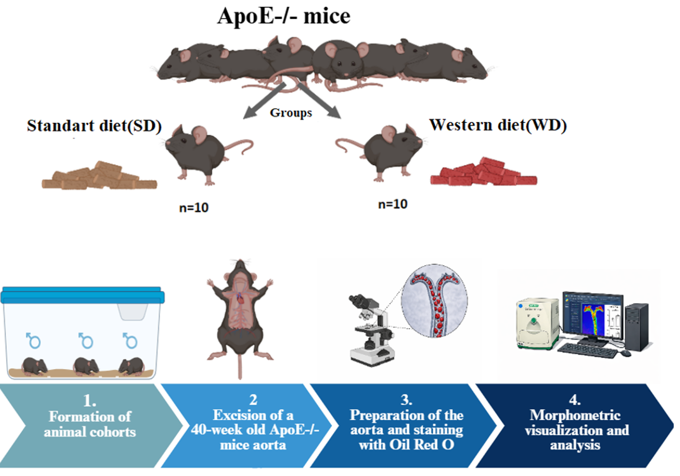
Materials and Methods: The study was performed on 20 male ApoE−/− mice (40 weeks old) divided into groups receiving a standard diet or a Western diet. After anesthesia, PBS perfusion was performed; the heart with the aorta was excised and fixed in 10% formalin; the aorta was cleaned and stained with Oil Red O. The aorta was longitudinally opened, photographed (GelDoc), and quantitatively analyzed using a Python script. Data are presented as mean ± SD; statistical significance was assessed using Student’s t-test at p < 0.05.
Results and Discussion: Thus, the use of the Oil Red O staining method in combination with automated image analysis in Python enables a transition from subjective assessment to precise quantitative measurement of the area of atherosclerotic lesions. This approach increases the reliability of screening therapeutic interventions in experimental models and enhances the statistical power of the study.
Conclusion: In this study, an original approach to morphometric visualization, analysis, and quantitative assessment of atherosclerotic lesions of the vascular wall in an apolipoprotein E–deficient mouse model (ApoE−/−) is proposed and validated for the first time.
Графическая аннотация
Ключевые слова:
atherosclerosis, aorta, ApoE−/− model, knockout mice, apolipoprotein E deficiency, morphometric analysisБиблиографические ссылки
Cao Q, Zhao J, Xing M, Xiao H, Zhang Q, Liang H, Ji A, Song S (2020) Current research landscape of marine-derived anti-atherosclerotic substances. Marine Drugs 18(9): 440. https://doi.org/10.3390/md18090440 [PubMed] [PMC]
Chen H, Howatt DA, Franklin MK, Amioka N, Sawada H, Daugherty A, Lu HS (2022) A mini-review on quantification of atherosclerosis in hypercholesterolemic mice. Global Translational Medicine 1(1): 72. https://doi.org/10.36922/gtm.v1i1.76 [PubMed] [PMC]
Daugherty A, Tall AR, Daemen MJAP, Falk E, Fisher EA, García-Cardeña G, Lusis AJ, Owens AP 3rd, Rosenfeld ME, Virmani R; American Heart Association Council on Arteriosclerosis, Thrombosis and Vascular Biology; and Council on Basic Cardiovascular Sciences (2017) Recommendation on design, execution, and reporting of animal atherosclerosis studies. Arteriosclerosis, Thrombosis and Vascular Biology 37(9): e131–e157.https://doi.org/10.1161/ATV.0000000000000062 [PubMed]
Evans NR, Bhakta S, Chowdhury MM, Markus H, Warburton E (2024) Management of carotid atherosclerosis in stroke. Practical Neurology 24(5): 382–386. https://doi.org/10.1136/pn-2023-003918 [PubMed]
Getz GS, Reardon CA (2016) Diet and murine atherosclerosis. Arteriosclerosis, Thrombosis and Vascular Biology 36(7): 1311–1318. https://doi.org/10.1161/01.ATV.0000201071.49029.17 [PubMed] [PMC]
He L, Chen Q, Wang L, Pu Y, Huang J, Cheng CK, Luo JY, Kang L, Lin X, Xiang L, Fang L, He B, Xia Y, Lui KO, Pan Y, Liu J, Zhang CL, Huang Y (2024) Activation of Nrf2 inhibits atherosclerosis in ApoE−/− mice through suppressing endothelial cell inflammation and lipid peroxidation. Redox Biology 74: 103229. https://doi.org/10.1016/j.redox.2024.103229 [PubMed] [PMC]
Hinder LM, Vincent AM, Hayes JM, McLean LL, Feldman EL (2013) Apolipoprotein E knockout as the basis for mouse models of dyslipidemia-induced neuropathy. Experimental Neurology 239: 102–110. https://doi.org/10.1016/j.expneurol.2012.10.002 [PubMed] [PMC]
Holmberg O, Lenz T, Koch V, Alyagoob A, Utsch L, Rank A, Sabic E, Seguchi M, Xhepa E, Kufner S, Cassese S, Kastrati A, Marr C, Joner M, Nicol P (2021) Histopathology-based deep learning predicts atherosclerotic lesions in intravascular imaging. Frontiers in Cardiovascular Medicine 8: 779807. https://doi.org/10.3389/fcvm.2021.779807[PubMed] [PMC]
Libby P, Buring JE, Badimon L, Hansson GK, Deanfield J, Bittencourt MS, Tokgözoğlu L, Lewis EF (2019) Atherosclerosis. Nature Reviews Disease Primers 5(1): 56. https://doi.org/10.1038/s41572-019-0106-z [PubMed]
Miano JM, Fisher EA, Majesky MW (2021) Fate and state of vascular smooth muscle cells in atherosclerosis. Circulation 143(21): 2110–2116. https://doi.org/10.1161/CIRCULATIONAHA.120.049922 [PubMed] [PMC]
Mohanta S, Yin C, Weber C, Hu D, Habenicht AJ (2016) Aorta atherosclerosis lesion analysis in hyperlipidemic mice. Bio-Protocol Journal 6(11): e1833. https://doi.org/10.21769/bioprotoc.1833 [PubMed] [PMC]
Noonan J, Cardoso L, Bobik A, Peter K (2025) Atherosclerotic plaque instability and rupture: recommended mouse models to empower clinically relevant discoveries, diagnostics, and therapeutics. Arteriosclerosis, Thrombosis and Vascular Biology 45(10): 1707–1714. https://doi.org/10.1161/ATVBAHA.125.321011 [PubMed] [PMC]
Pedro-Botet J, Climent E, Benaiges D (2020) Atherosclerosis and inflammation: New therapeutic approaches. Medicina Clinica 155(6): 256–262. https://doi.org/10.1016/j.medcli.2020.04.024 [PubMed]
Shcheblykin DV, Bolgov AA, Pokrovskii MV, Stepenko JV, Tsuverkalova JM, Shcheblykina OV, Golubinskaya PA, Korokina LV (2022) Endothelial dysfunction: developmental mechanisms and therapeutic strategies. Research Results in Pharmacology 8(4): 115–139. https://doi.org/10.3897/rrpharmacology.8.80376
Sheng R, Li YN, Wang L, Jiang XH, Xiao TM, Zhang YH, Li SW, Wu YX, Xu Y, Xu YN, Si SY (2024) A novel small-molecule PCSK9 inhibitor E28362 ameliorates hyperlipidemia and atherosclerosis. Acta Pharmacologica Sinica 45(10): 2119–2133. https://doi.org/ 10.1038/s41401-024-01305-9 [PubMed] [PMC]
Stadelmann VA, Boyd G, Guillot M, Bienvenu JG, Glaus C, Varela A (2021) Automatic quantification of atherosclerosis in contrast-enhanced microCT scans of mouse aortas ex vivo. International Journal of Biomedical Imaging 2021: 4998786. https://doi.org/10.1155/2021/4998786 [PubMed] [PMC]
Stone PH, Libby P, Boden WE (2023) Fundamental pathobiology of coronary atherosclerosis and clinical implications for chronic ischemic heart disease management –the plaque hypothesis: a narrative review. JAMA Cardiology 8(2): 192–201. https://doi.org/10.1001/jamacardio.2022.3926 [PubMed] [PMC]
Загрузки
Дополнительные файлы
Опубликован
Как цитировать
Выпуск
Раздел
Лицензия
Copyright (c) 2026 Lebedev PR, Syrykh NV, Pokrovskii MV

Это произведение доступно по лицензии Creative Commons «Attribution» («Атрибуция») 4.0 Всемирная.
 Русский
 English

