Effective wound healing agents based on N-alkenylimidazole zinc complexes derivatives: future prospects and opportunities
DOI:
https://doi.org/10.18413/rrpharmacology.9.10047Аннотация
Introduction: The therapeutic effect of commercially available domestic and foreign drugs for the treatment of various skin injuries is far from optimal. These drugs have no universal effects, but cause pronounced side reactions. There is a clear demand for development of innovative wound-healing drugs with antimicrobial properties, which increase the natural protective function of the skin. Pharmaceutical compounds with zinc nanoparticles have been increasingly recognized as a promising therapeutic direction. These drugs can easily penetrate into damaged tissues and stimulate metabolic processes. Zinc complexes with imidazole derivatives are of a particular interest. Imidazole acts as a structural fragment of many natural physiologically active compounds, thus providing targeted delivery of this essential trace element into the wound for inclusion in the multicascade mechanism of wound healing. The aim of the study: to provide experimental evidence for effects of recently developed zinc complexes with N-alkenylimidazole as wound healing agents.
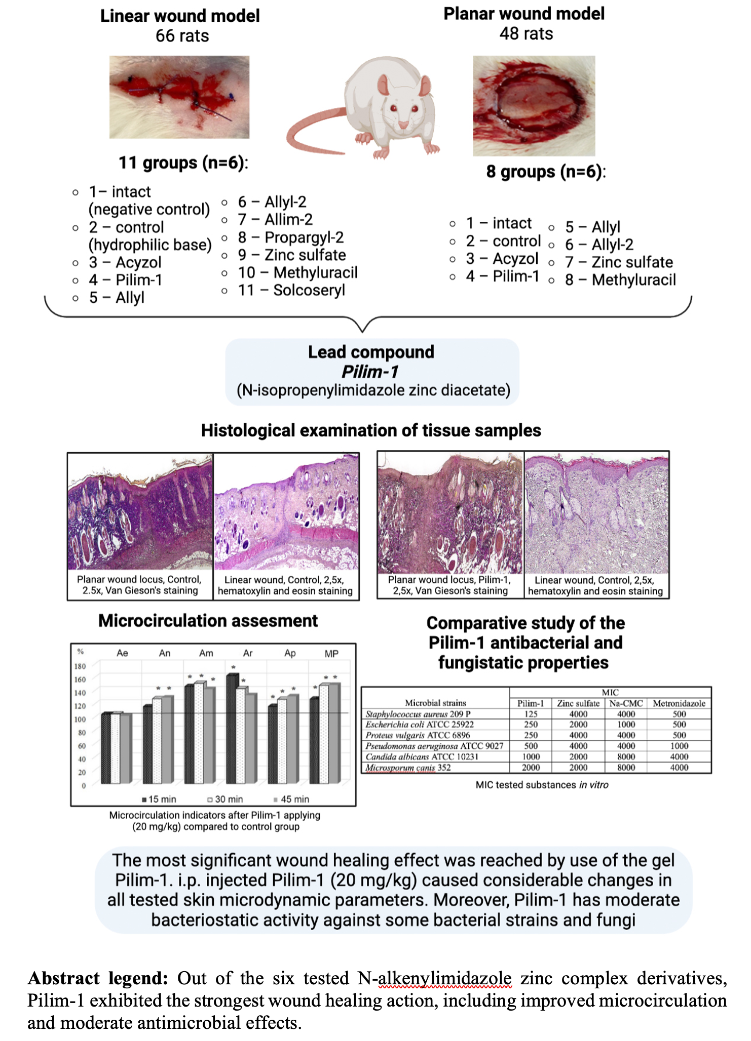
Materials and Methods: Wound-healing effects of six 1% gels containing distinct N-alkenylimidazole zinc complexe derivatives based on the Na-carboxymethylcellulose (Na-CMC) were comparatively studied in 128 outbred white rats of both genders. The Na-CMC-based Zinc Sulfate 1% gel, Methyluracil and Solcoseryl served as reference drugs. After performing the local tolerance study of zinc complexes, linear and planar sterile wounds of comparable size were inflicted in anesthetized animals. The degree of healing was evaluated on the day 8 and day 28 after the treatment start by wound sizes and histological examination of inflammatory response, epithelization, granulation tissue, angiogenesis, and necrosis. The skin microcirculation system was evaluated using the laser Doppler Flowmetry (LDF), whereby the blood flow indicators were recorded 30 and 60 minutes after intraperitoneal administration of the trial compound. The antimicrobial activity of the zinc compounds was determined in vitro by means of their minimum inhibitory concentration suppressing the bacteria and fungi growth using the double serial dilution method in liquid culture media. The statistical data processing was performed using the Statistica 12 software package.
Results and Discussion: In the linear wound model, all animals treated with either of six experimental zinc compounds showed almost complete reduction in wound size (92-100%, p<0.05) on the day 8, significantly exceeding the wound healing in the control animals (reduction by 67-88 %, p<0.05) and effects of the reference drugs (reduction by 83-86%, p<0.05). In the planar wound model, the most significant wound healing effect was reached by using the gel containing N-isopropenylimidazole zinc diacetate (encoded as Pilim-1). The respective histological examination showed signs of complete epithelialization, absence of destructive changes in the epidermis, restoration of skin appendages and presence of mature granulation tissue. Intraperitoneal Pilim-1 administration at a dose of 20 mg/kg improved microcirculation in the rat skin, as judged by significant effects on perfusion and the amplitudes of the isolated rhythms of the LDF-gram. In addition, Pilim-1 exerted a moderate bacteriostatic and fungistatic activity, which was 2 times greater than the antimicrobial activity of Metronidazole.
Conclusion: Topical application of gels containing 1% N-alkenylimidazole zinc complex derivatives accelerates the healing of uninfected linear and planar wounds in comparison with the established reference drugs. The Pilim-1 zinc compound exhibited the most pronounced therapeutic effect. The observed in vitro antimicrobial action of Pilim-1 is of further interest for potential implications in treatment of infected skin wounds. The regenerative effect of this substance opens prospects for development of new drugs with improved wound healing properties.
Графическая аннотация

Ключевые слова:
linear wound, N-alkenylimidazole derivatives, reparative regeneration, skin, planar wound, topical therapy, wound healing, zincБиблиографические ссылки
Atayik MC, Çakatay U (2023) Redox signaling in impaired cascades of wound healing: promising approach. Molecular Biology Reports 50(8): 6927–6936. https://doi.org/10.1007/s11033-023-08589-w [PubMed]
Aliev G, Li Y, Chubarev VN, Lebedeva SA, Parshina LN, Trofimov BA, Sologova SS, Makhmutova A, Avila-Rodriguez MF, Klochkov SG, Galenko-Yaroshevsky PA, Tarasov VV (2019) Application of acyzol in the context of zinc deficiency and perspectives. International Journal of Molecular Sciences 20 (9): 2014. https://doi.org/10.3390/ijms20092104 [PubMed] [PMC]
Adrianova II, Kolesnik VM, Galkina OP, Ostrovsky AV (2016) Treatment of erosive lesions of the oral mucosa with using solkoseril dental adhesive paste. Tauride Medical and Biological Bulletin [Tavricheskiy Mediko-Biologicheskiy Vestnik] 19 (1): 5–7. [in Russian]
Baikalova LV, Sokol VI, Khrustalev VN, Zel’bst EA, Trofimov BA (2005) Crystal and molecular structure of bis(1-vinylimidazole)diacetatozinc. Russian Journal of General Chemistry [Zhurnal Obshhei Khimii] 75(9): 1542–1547. https://doi.org/10.1007/s11176-005-0448-y [in Russian]
Barinov AV, Nechiporenko SP (2006) Developed carbon monoxide antidote. UNIFOR RASHA 2006: 116–117 [in Russian]
Bunnell BE, Escobar JF, Bair KL, Sutton MD, Crane JK (2017) Zinc blocks SOS-induced antibiotic resistance via inhibition of RecA in Escherichia coli. PLoS ONE 12(5): e0178303. https://doi.org/10.1371/journal.pone.0178303[PubMed] [PMC]
Cheknov SB, Vostrova EI, Apresova MA, Piskovskaya LS, Vostrov AV (2015) Inhibition of bacterial growth in the cultures of Staphylococcus aureus and Pseudomonas aeruginosa in the presence of copper and zinc cations. Journal of Microbiology, Epidemiology and Immunobiology [Zhurnal Mikrobiologii, Epidemiologii i Immunobiologii] 2: 9–17. [in Russian]
Cheknov SB, Vostrova EI, Sarycheva MA, Kisil SV, Anisimov VV, Vostrov AV (2017) Inhibition of bacterial growth in the cultures of Streptococcus pyogenes and Streptococcus agalactiae in the presence of copper and zinc cations. Journal of Microbiology, Epidemiology and Immunobiology [Zhurnal Mikrobiologii, Epidemiologii i Immunobiologii] 3: 26–35. [in Russian]
Checknov SB, Vostrova EI, Sarycheva MA, Vostrov AV (2019) Protective effects of zinc cations against Staphylococus aureus exposed to antibiotics. Journal of Microbiology, Epidemiology and Immunobiology [Zhurnal Mikrobiologii, Epidemiologii i Immunobiologii] 6: 5–12. [in Russian]
El-Adl M, Abdelkhalek N, Mahgoub HA, Salama MF, Ali M (2018) Improved healing of the deeply incisional wounds in partially scaled common carp by zinc sulphate bath. Aquaculture Research 49(10): 3411–3420. https://doi.org/10.1111/are.13805
Grigoryan AY, Gorokhova AS, Belozerova AV (2017) Wound coating with chlorhexidine and metronidazol in the treatment of wounds. [Byulleten` Severnogo Gosudarstvennogo Meditsinskogo Universiteta] 21(37): 4–5. [in Russian]
Krarup P-M, Eld M, Jorgensen LN, Hansen MB, Agren MS (2017) Selective matrix metalloproteinase inhibition increases breaking strength and reduces anastomotic leakage in experimentally obstructed colon. International Journal of Colorectal Disease 32(9): 1277–1284. https://doi.org/10.1007/s00384-017-2857-x [PubMed]
Krishnaswamy VR, Mintz D, Sagi I (2017) Matrix metalloproteinases: The sculptors of chronic cutaneous wounds. Biochimica et Biophysica Acta. Molecular Cell Research 1864(11): 2220–2227. https://doi.org/10.1016/j.bbamcr.2017.08.003 [PubMed]
Krupatkin AI (2018) The role of oscillatory processes in the diagnosis of the state of microcirculatory tissue systems. Physiology of Man [Fiziologiya Cheloveka] 44 (5): 103–114. https://doi.org/10.1134/S0131164618050077 [in Russian]
Krupatkin AI, Sidorov VV (2005) Laser Doppler Flowmetry of blood microcirculation. Meditsina, Moscow, 254 pp. [in Russian]
Larsen H, Ahlström MG, Gjerdrum LMR, Mogensen M, Ghathian K, Calum H, Ågren MS (2017) Noninvasive measurement of reepithelialization and microvascularity of suction-blister wounds with benchmarking to histology. Wound Repair and Regeneration 25(6): 984–993. https://doi.org/10.1111/wrr.12605 [PubMed]
Lebedeva SA, Galenko-Yaroshevsky PA (Jr.), Samsonov MYu, Erlich AB, Margaryan AG, Materenchuk MYu, Arshinov IaR, Zharov YuV, Zelenskaya AV, Shelemekh OV, Lomsadze IG, Demura TA (2023) Molecular mechanisms of wound healing: the role of zinc as an essential microelement. Research Results in Pharmacology 9(1): 25–39. https://doi.org/10.18413/rrpharmacology.9.10003
Lin P-H, Sermersheim M, Li H, Lee PHU, Steinberg SM, Ma J (2017) Zinc in wound healing modulation. Nutrients 10(1): 16. https://doi.org/10.3390/nu10010016 [PubMed] [PMC]
Mironov AN (2012) Guidelines for conducting preclinical drug trials. Griff and K, Moscow, 944 pp. [in Russian]
Nozdrin VI, Belousova TA, Iatskovskiĭ AN (2002) Morphological aspects of dermatotrophic action of methyluracil applied epicutaneously. Morfologiia 122(5): 74–78. [PubMed]
Ogai MA, Stepanova EF, Dzyuba VF, Morozova EV (2010) The use of polymer bases in ointments for the treatment and prevention of diabetic foot pathology. Scientific Journal of BelSU. Medicine, Pharmacy [Nauchnye vdomosti BelGU. Meditsina, Farmatsiya] 22(93): 5–9. [in Russian]
Parchina LN, Grishcenko LA, Smirnov VI, Borodina TN, Shakhmardanova SA, Tarasov VV, Apartsin KA, Kireeva VV, Trofimov BA (2019) Synthesis, characterization and biological evaluation of Zn (II) and Co (II) complexes of N-allylimidazole as potential hypoxia-targeting agents. Polyhedron 161: 126–131. https://doi.org/10.1016/j.poly.2019.01.005
Raghunath A, Perumal E (2017) Metal oxide nanoparticles as antimicrobial agents: a promise for the future. International. Journal of Antimicrobial 49(2): 137–152. https://doi.org/10.1016/j.ijantimicag.2016.11.011 [PubMed]
Roy S, Khanna S, Nallu K, Hunt TK, Sen CK (2006) Dermal wound healing is subject to redox control. Molecular Therapy 13: 211–220. https://doi.org/10.1016/j.ymthe.2005.07.684 [PubMed] [PMC]
Shakhmardanova SA, Babaniyazova ZH, Tarasov VV, Pevnev GO, Chubarev VN, Sologova SS (2017) Protective effect of acyzol in a model of carbon tetrachloride-induced hepatotoxicity. BioNanoScience 7(2): 329–332. https://doi.org/10.1007/s12668-016-0352-4
Shakhmardanova SA, Zelenskaya AV, Galenko-Yaroshevsky PA (2016) Metal complexes based on the N-alkenilimidazol as redox-regulators of hypoxic conditions. Journal of Fundamental Medicine and Biology [Zhurnal Fundamental`noi Meditsiny i Biologii] (3): 63–67. [in Russian]
Tiganov SI, Grigoryan AI, Blinkov YU, Pankrusheva TA, Mishina EU, Giliaeva LV (2018) The use of miramistin and metronidazole in the treatment of experimental purulent wounds. Siberian Medical Review [Sibirskoe Meditsinskoe Obozrenie] (1): 43–48. https://doi.org/10.20333/2500136-2018-1-43-48 [in Russian]
Tiwari V, Mishra N, Gadani K, Solanki PS, Shah NA, Tiwari M (2018) Mechanism of anti-bacterial activity of zinc oxide nanoparticle against carbapenem-resistant Acinetobacter Baumannii. Frontiers in Microbiology 9: 1218. https://doi.org/10.3389/fmicb.2018.01218 [PubMed] [PMC]
Xie Y, He Y, Irwin PL, Jin T, Shi X (2011) Antibacterial activity and mechanism of action of zinc oxide nanoparticles against campylobacter jejuni. Applied and Environmental Microbiology 77(7): 2325–2331. https://doi.org/10.1128/AEM.02149-10 [PubMed] [PMC]
Yakimoskii AF, Shantyr II, Vlasenko MA, Yakovleva MV (2017) Effects of acyzol on zinc content in rat brain and blood plasma. Bulletin of Experimental Biology and Medicine 162(3): 293–294. https://doi.org/10.1007/s10517-017-3597-1 [PubMed]
Загрузки
Опубликован
Как цитировать
Выпуск
Раздел
Лицензия
Copyright (c) 2023 Svetlana A. Lebedeva, Pavel A. Galenko-Yaroshevsky (Jr.), Tatiana V. Fateeva, Sergey S. Pashin, Nataliya R. Pashina, Irina B. Nektarevskaya, Andrey V. Zadorozhniy, Olga V. Shelemekh, Marina Yu. Ravaeva, Elena N. Chuyan, Lusine O. Alukhanyan, Tereza R. Glechyan, Kerim Mutig, Maria Yu. Materenchuk

Это произведение доступно по лицензии Creative Commons «Attribution» («Атрибуция») 4.0 Всемирная.
 Русский
 English

